Tourmaline in Cheviot Igneous Rocks
Tourmaline occurs as a secondary mineral in highly altered Cheviot rocks appearing as both prismatic and acicular crystals between 1 to 2.5 mm in length that are pleochroic from yellow, through purplish brown to dark blue and blue-green and sometimes showing evidence of zoning. The crystals can occur in a radiating texture that may reflect their formation as late-stage replacement of biotite, plagioclase and microperthite.
Al-Hafdh analysed fourteen grains of tourmaline by electron microprobe from three different samples of medium to fine-grained porphyritic rock (his Linhope, and Hedgehope varieties) and one sample of evolved granite (his Woolhope variety).
A single tourmaline grain from one of the fine-grained porphyritic samples was scanned to chart changes in composition from core to rim.
The first three samples proved to be of very similar composition all sharing relatively high proportions of calcium and magnesium. Al-Hafdh thought their composition reflected the chemical composition of the fluids associated with the sericitic and propylitic processes that altered these rocks.
The analysis of the zoned crystal revealed a compositional change from a relatively magnesium, calcium and titanium rich core to a low magnesium rim that was also depleted in calcium. This variation was thought to reflect the changing chemical composition of the hydrothermal fluids.
Plotting relative proportions of iron, magnesium and aluminium reveals a composition intermediate between schorl and dravite.
Composition Diagram for Cheviot Tourmaline

Identification
Tourmaline has no cleavage, only irregular cracks in the crystals. It has moderately high birefringence although its second order colours are masked by its characteristically strong pink, purple-brown to blue-green absorption colour that clearly changes as the crystal is rotated on the microscope stage in plane polarised light.
It has a moderately high refractive index so the Becke line moves into a tourmaline crystal that is adjacent to a quartz or feldspar crystal when the stage is moved down, away from the objective lens.
A prismatic tourmaline crystal will show parallel extinction when viewed with crossed polarising filters.
In PPL, shorl tends to show grey, blue, green or pink colours and dravite tends to appear yellow and pale brown.
Photomicrographs of Cheviot Tourmaline

Both of these poorly formed tourmaline crystals exhibit zoning and quartz inclusions.

Locality: The Cheviot.
The prismatic tourmaline crystal in the centre of view is positioned so that it’s long axis is parallel to the vibration direction of the transmitted plane polarised light. In this position it reveals its cleanest/lightest absorption colour, a light pinkish brown.

In this position its absorption colour is a darker purple-brown.


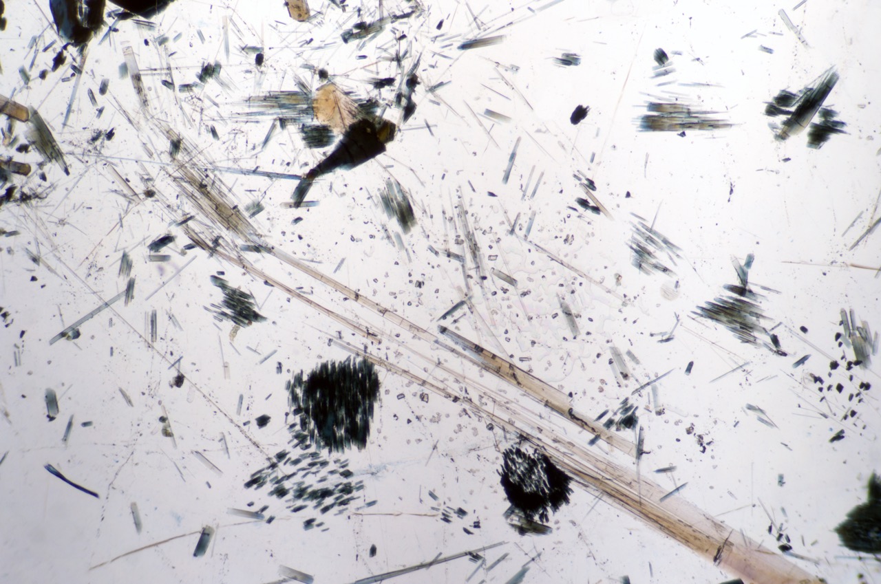
Here tourmaline takes on an acicular form i.e. the form of needle-like structures.