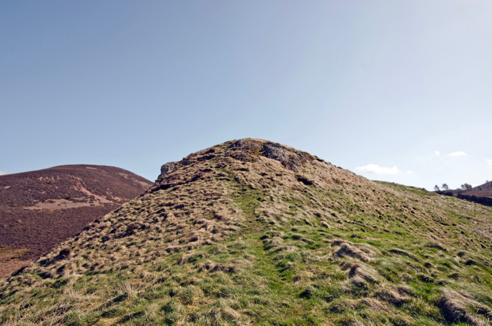
Location 1. Little Hill summit basalt outcrop at NT546319
Location 2. Little Hill lower basalt outcrop at NT546319
Location 3. Lower Wester Hill rhyolite exposure at NT548318
Location 4. Wester Hill summit rhyolite exposure at NT548315
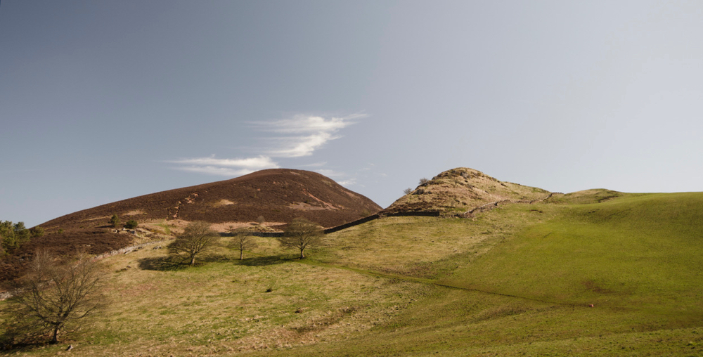
The Eildon Hills rise just to the south of the attractive Roxburghshire town, Melrose. There are three sizable hills of igneous material; Mid Hill, Wester Hill,and North Hill, and a smaller fourth one aptly named, Little Hill – all surrounded by a fringe of Old Red Sandstone rock that has been eroded away elsewhere but has been protected here by the harder igneous rock. The three larger hills are thought to be the revealed remains of a composite laccolith that was intruded into the Upper Old Red Sandstone sedimentary strata about 352 million years ago in the early Carboniferous.
The hills were the subject of a detailed study in 1914 by the renowned geologist Lady Rachel McRobert and much that has been written since relies heavily on her work. She identified six distinct layers to the intrusion and, noting the spatial and angular relationships of the layers on Mid Hill and Wester Hill along with the positions and similar composition of spurs on the western side of the hills together with dykes in the vicinity, suggested an original flat-bottomed, domed-topped ‘laccolithic’ form of a much greater mass than that suggested by the three main hills as they stand. McRobert’s beautifully drawn original maps and cross-section clearly communicate her findings and hypotheses.
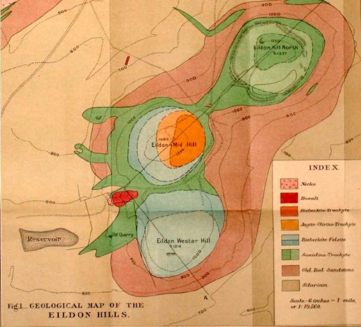

Wester Hill has a partial base-layer of porphyritic sanidine-trachyte below two distinct layers of what McRobert terms riebeckite-felsite, but classified more recently as riebeckite-rhyolite or, according to other sources, simply as rhyolite. The latter is in keeping with our finding no riebeckite in any of our samples of the Eildon rocks apart from one sample of trachyte from upper Mid Hill.
Mid Hill has the same sanidine-trachyte base and felsite/rhyolite layers as Wester Hill but with an additional layer of augite-olivine-trachyte and an upper layer of riebeckite-trachyte.
North Hill shares the same porphyritic sanidine-trachyte base below a layer of non-porphyritic sanidine-trachyte and an upper layer of porphyritic sanidine-trachyte.
Little Hill has been identified as a later intrusion than its larger cousins and has been described as a small basalt-plugged vent with associated agglomerate and as a neck that originally fed a volcano that once sat on top of the laccolith, since eroded away.
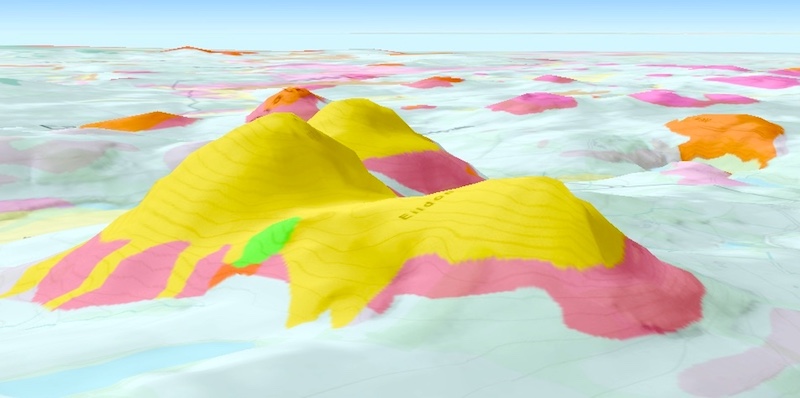
Exaggerated 3D view from the south-west from the BGS online geology map.
There are two easy routes into the Eildons, that from the north via the footpath by the golf course and the route from the west via a footpath leading from the B6359 road. We choose the latter and park on the raised verge near the gated track leading to the Bowdenmoor quarry. The footpath, just to the south of here, leads through a field gate and across rough, waterlogged ground to two wicket gates below Little Hill.
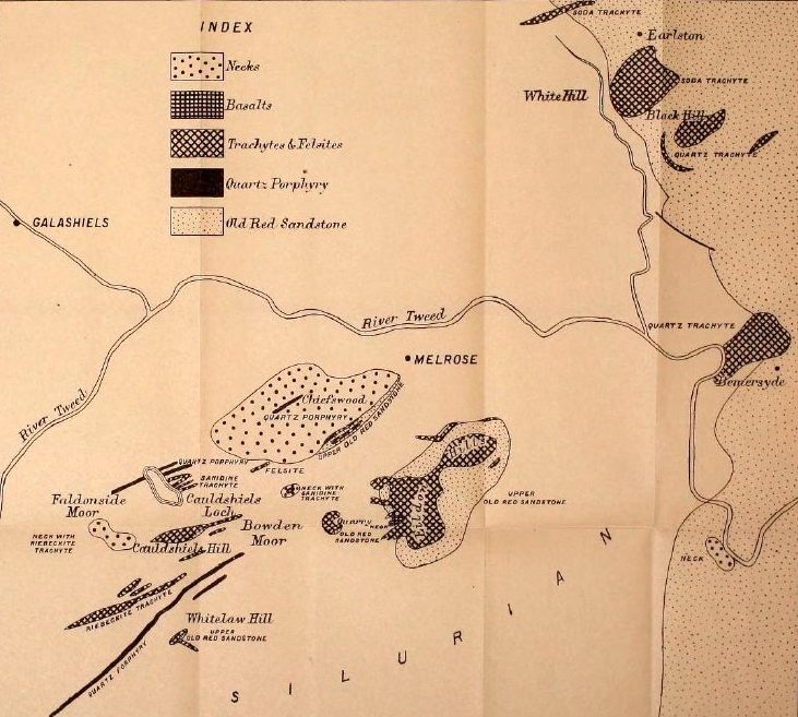
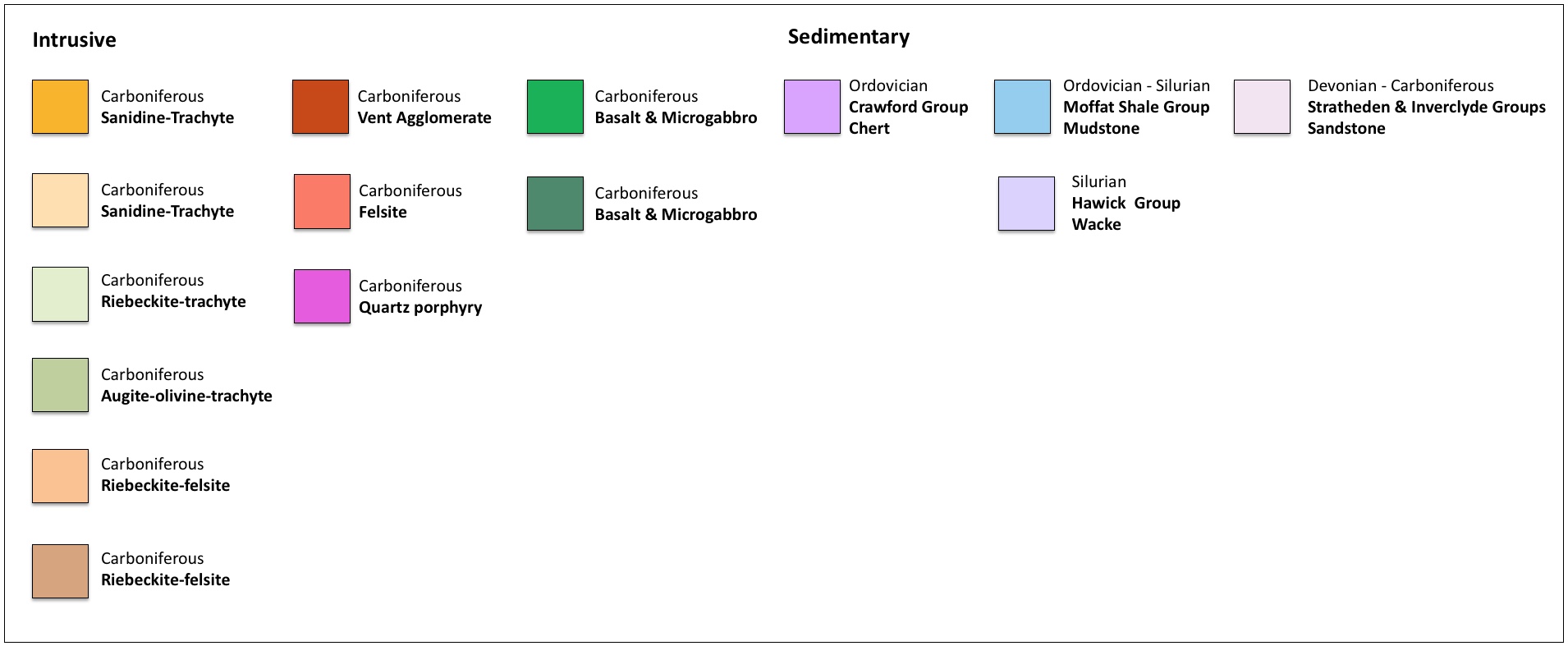
Map showing locations and rock types around the Eildon Hills

Locations 1. Little Hill summit basalt outcrop

Our first locations are the upper and lower exposures of basalt on Little Hill. We find the rock at both is very weak and crumbly. That at the summit is finer-grained and without phenocrysts but with some amygdales while the lower one, on the west slope, is said to be more of a ‘Markle’ type of basalt with some plagioclase phenocrysts as well as the occasional remains of olivine phenocrysts.
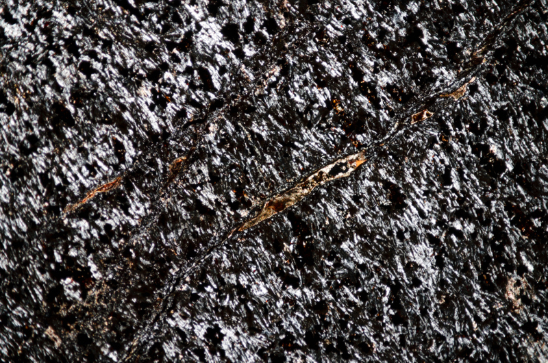
In thin section we don’t see the any obvious signs of altered olivine but in the summit basalt, we do see the flow patterns of the plagioclase laths, masses of iron-titanium oxides, carbonate-filled amygdales, occasional small flakes of biotite, and one example of a slightly pleochroic crystal with first to second order interference colours and an inclined extinction, perhaps an amphibole, embedded in a patch of carbonate material.
An explanation for the presence of basalt at this location posits a mass of basaltic magma deep in the crust that stratified following fractional crystallisation with more felsic and less dense trachytic and rhyolitic magma lying above the more mafic, denser basalt. The upper, felsic material was the first to move up towards the surface to be intruded as the laccolith and only later did the basaltic magma rise to be intruded as the neck at Little Hill and possibly erupted at the surface.
Agglomerate associated with this neck or vent is shown on maps to the south of the western spur of the hill, but we don’t investigate it on this occasion.
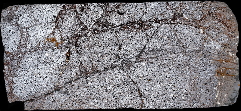
First sample
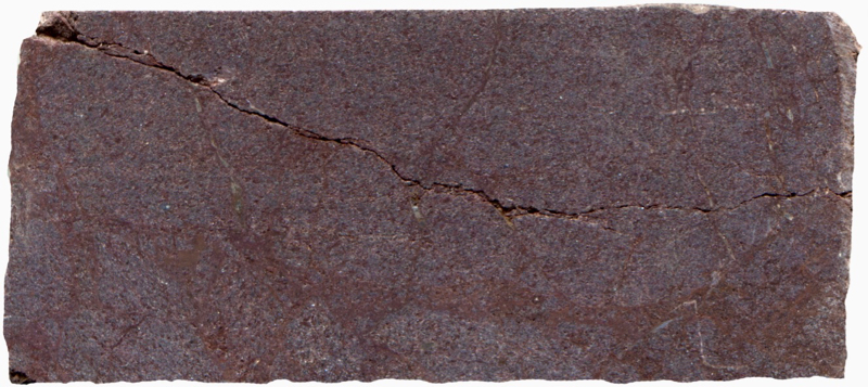
Prepared specimen viewed in plain reflected light (measures 42mm across)
The crumbly condition of the rock is evident in the thin section.
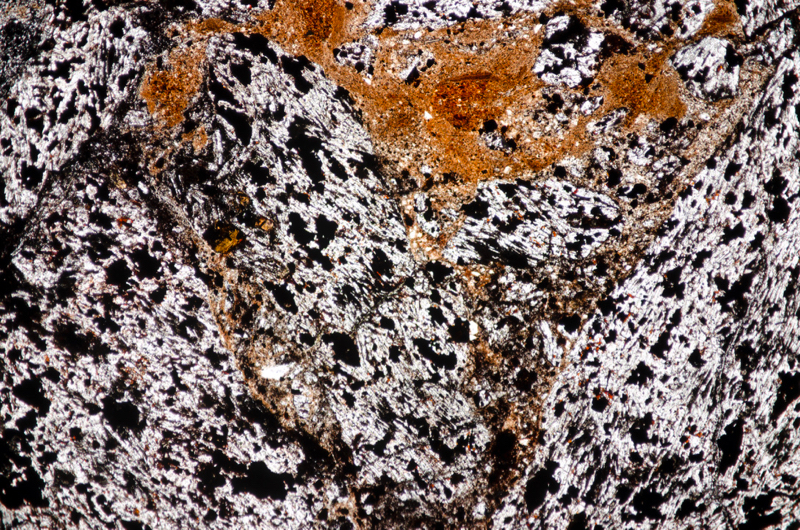
The rock appears to have been fractured early on resulting in the juxtaposition of volumes with different directions of magma flow. Section viewed in plane polarised light (FoV 4.6 x 3.0 mm)

Note the high proportion of primary iron-titanium oxides and the addition of later, orange-brown secondary oxides due to alteration.
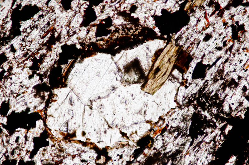
Perhaps these minerals represent an altered olivine phenocryst. Section viewed in plane polarised light (FoV 1.2 x 0.8 mm)
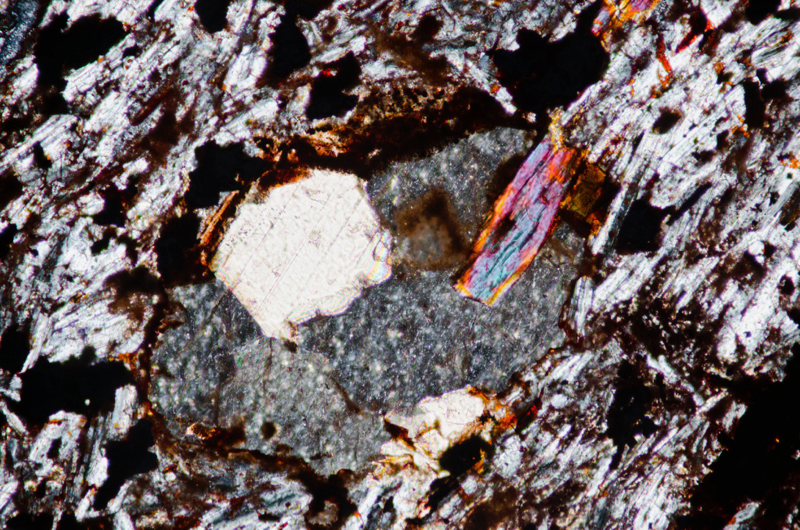
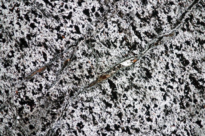
Section viewed in plane polarised light (FoV 4.6 x 3.0 mm)
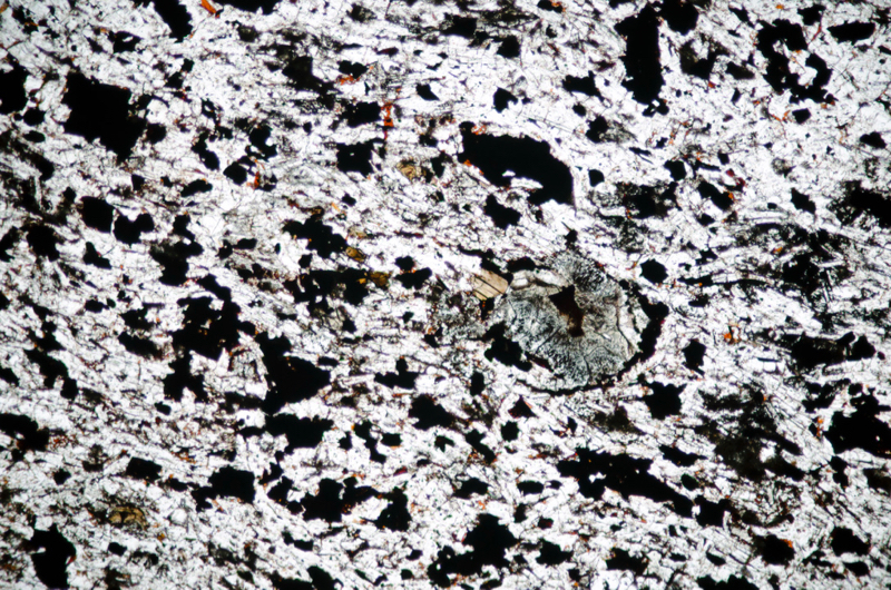
Section viewed in plane polarised light (FoV 2.3 x 1.5 mm)

The small flake of biotite, probably a secondary mineral, is at the very centre of the image.
Second sample
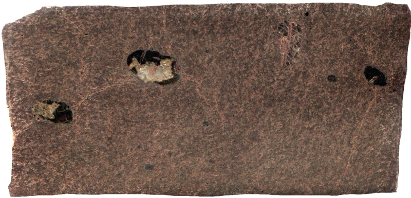
Prepared specimen viewed in plain reflected light (measures 42mm across)
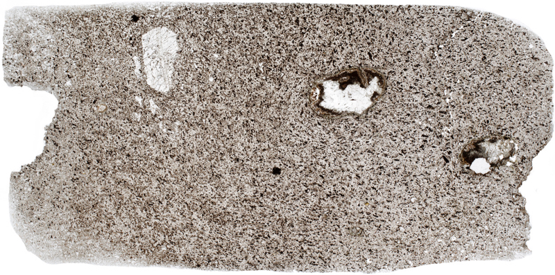


Section viewed with crossed polarising filters (FoV 4.6 x 3.0 mm)

Section viewed in plane polarised light (FoV 1.2 x 0.8 mm)
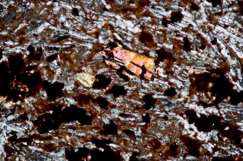
Location 2. Little Hill lower basalt outcrop
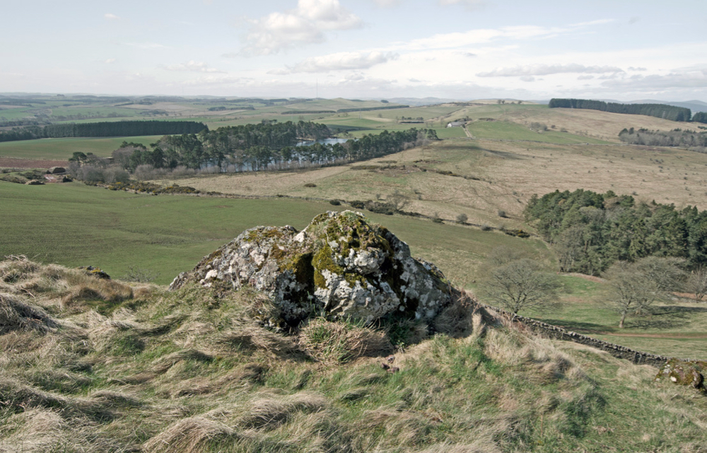
We find no amygdales in this basalt but we do see occasional plagioclase phenocrysts and granular pyroxene.
First sample
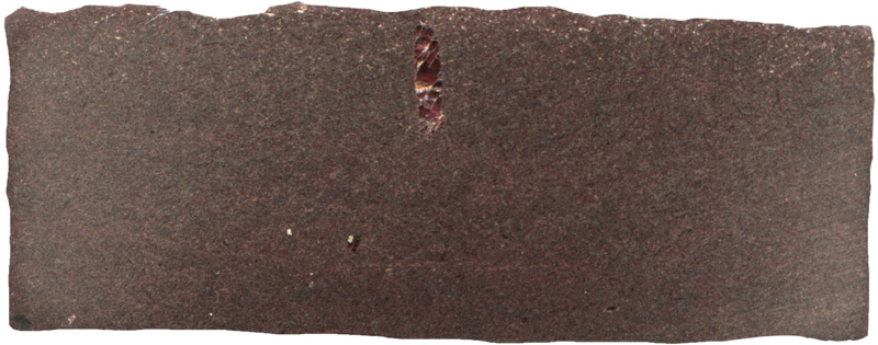
Prepared specimen viewed in plain reflected light (measures 42mm across)
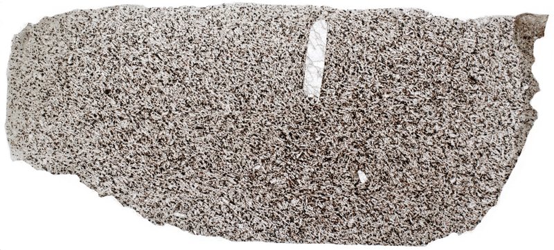
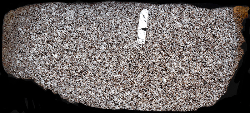

Section viewed with crossed polarising filters (FoV 4.6 x 3.0 mm)
Second sample
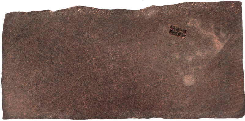
Prepared specimen viewed in plain reflected light (measures 42mm across)
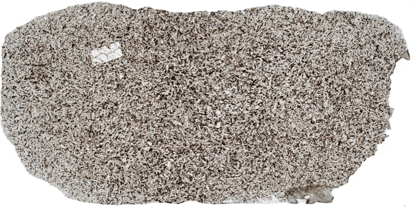

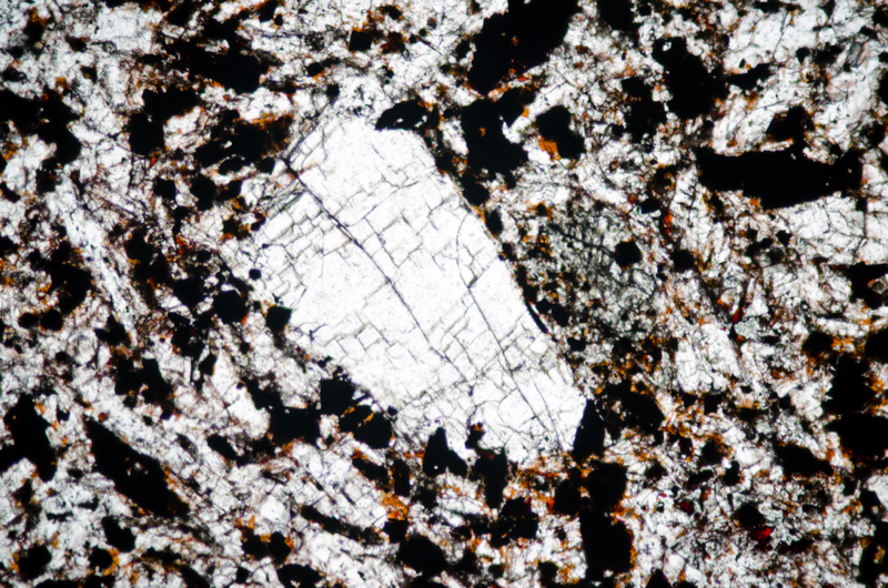
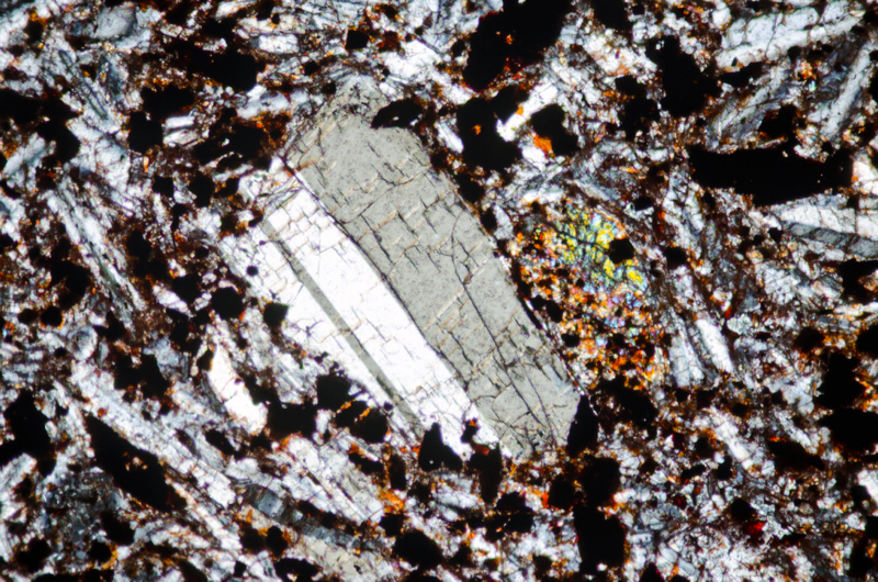
Section viewed in plane polarised light (FoV 2.3 x 1.5 mm)
Location 3. Lower Wester Hill rhyolite exposure at NT548318
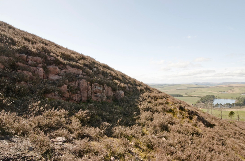
Our next locations are on Wester Hill. McRobert identified all of Wester Hill’s rock as riebeckite-felsite occurring in two distinct layers, the lower one being a pink rock, ‘with small dark patches of riebeckite. Under the microscope the riebeckite-growths are minute, and largely altered to limonite.’ In recent maps Wester Hill’s rock is classified as rhyolite.
From Little Hill, we take the path leading between Little Hill and Mid Hill and then follow the first path on the right up Wester Hill that takes us by the exposure of the lower layer of rhyolite that is shown above.
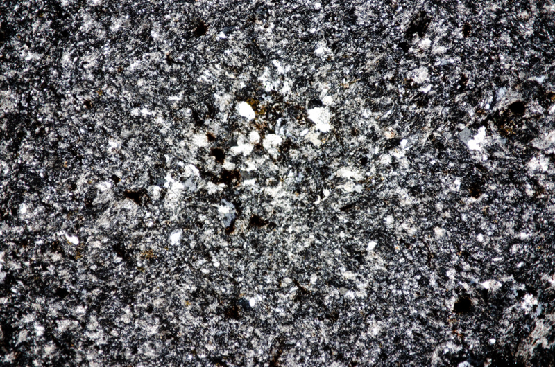
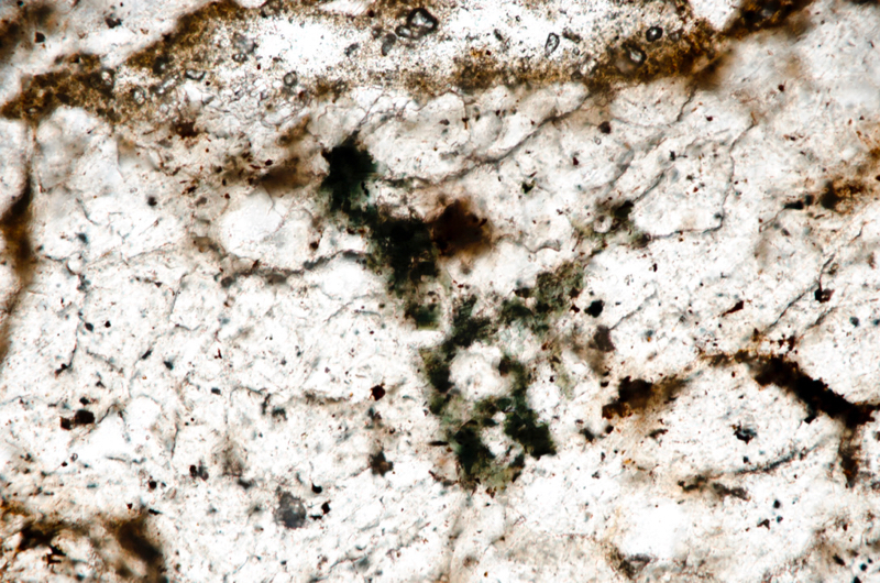
In the hand, we take the dark speckles in the rhyolite to be riebeckite but in thin section, it is clear that it is a form of biotite that has a light yellow-brown to very dark green-brown pleochroism, typical interference colours and parallel extinction. We find no riebeckite in our sample.
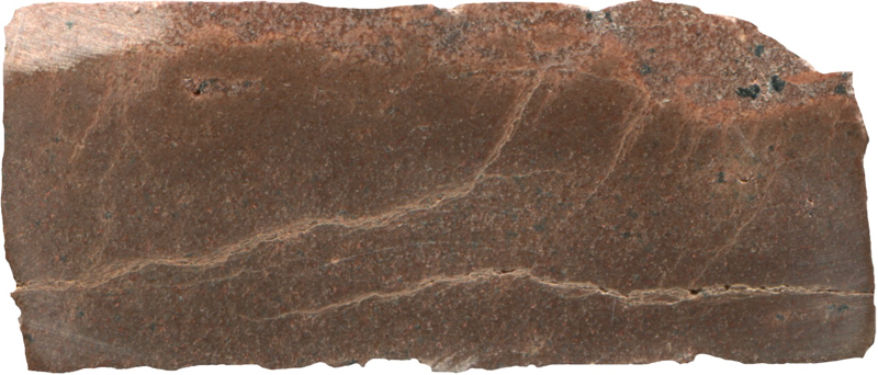
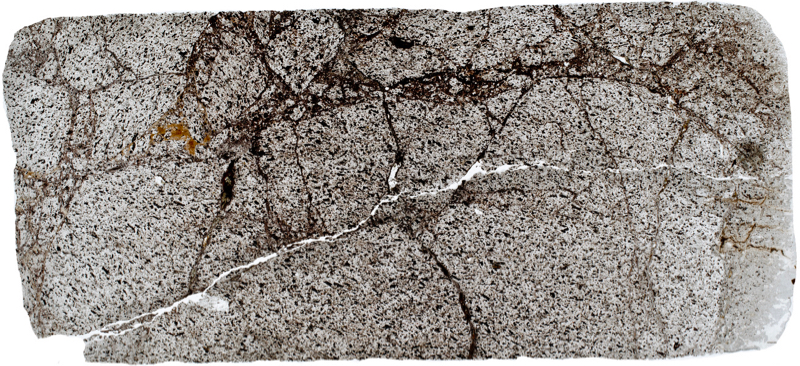
Prepared specimen viewed in plain reflected light (measures 42mm across)

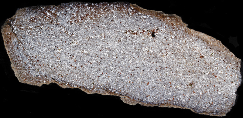
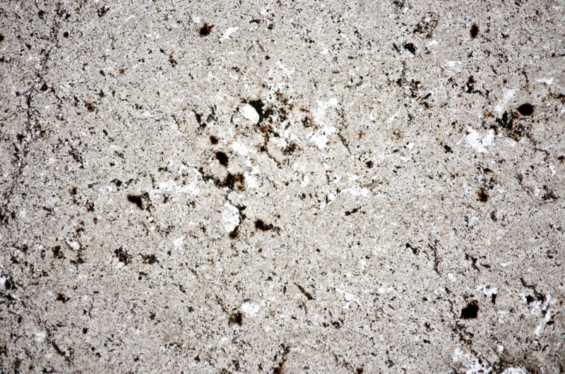
Section viewed in plane polarised light (FoV 4.6 x 3.0 mm)


Section viewed in plane polarised light (FoV 0.5 x 0.3 mm)
Location 4. Wester Hill summit rhyolite exposure at NT548315
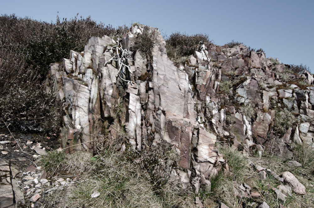
Note the size of the grass and bracken for scale.
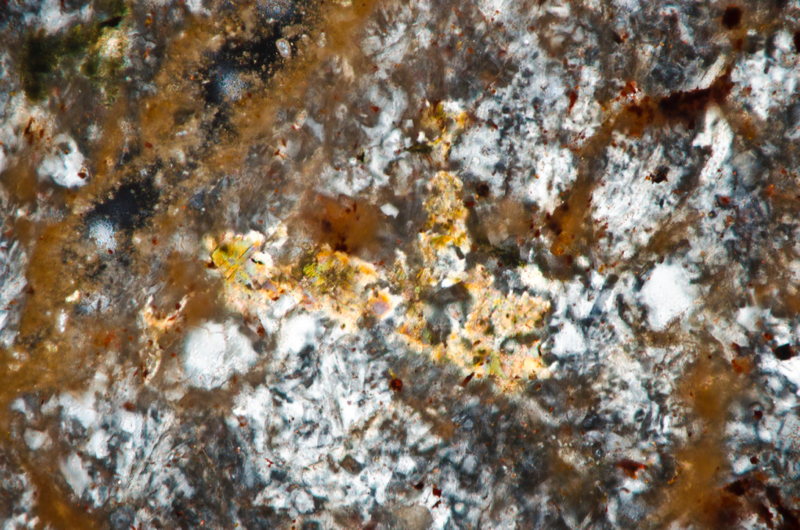
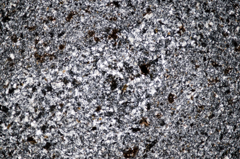
We continue to the summit where there are a number of small exposures of a particularly hard and splintery rhyolite that lies all around in shattered plates and forms the scree slopes on the southern side of the hill.
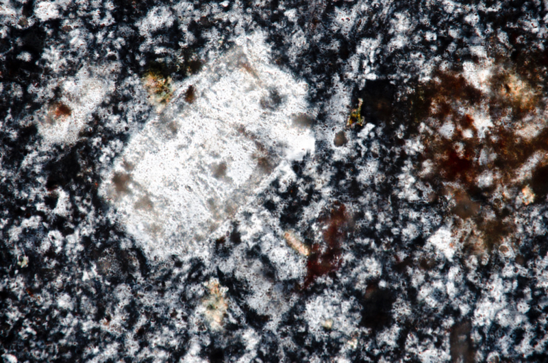
McRobert writes of this rock, ‘fresh riebeckite is abundant in nests and irregular aggregates: it is pleochroic. from deep blue to lemon-yellow. The ground-mass is felsitic, with much secondary quartz in large plates. The higher parts of the upper layer become coarser, and show microporphyritic sanidine-crystals ; a brownish-green biotite also occurs in one slide from the summit of the Wester Hill.’ She adds, ‘in these higher horizons there is an increasing proportion of soda, and phenocrysts of soda-orthoclase have been found in the corresponding rocks of the Mid Hill.’ Our sample from the summit appears less coarse than the rhyolite we found lower down.Again, riebeckite is absent but we do see it contains the occasional sanidine phenocryst along with more of the biotite.
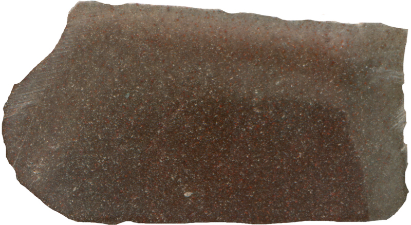
Prepared specimen viewed in plain reflected light (measures 42mm across)

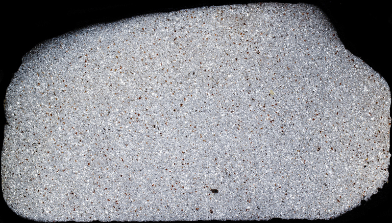

Section viewed in plane polarised light (FoV 4.6 x 3.0 mm)
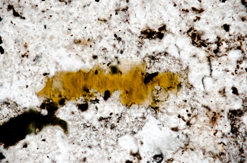
Section viewed in plane polarised light (FoV 0.5 x 0.3 mm)
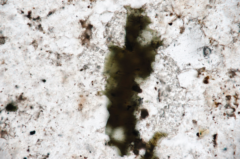
Section viewed in plane polarised light with slide rotated 90deg. to show pleochroism.(FoV 0.5 x 0.3 mm)
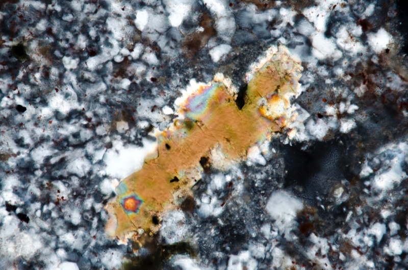
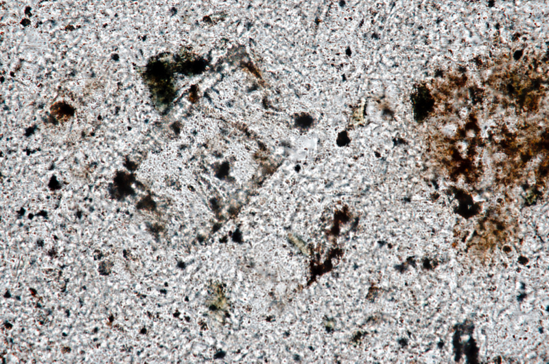
Section viewed in plane polarised light (FoV 1.2 x 0.8 mm)
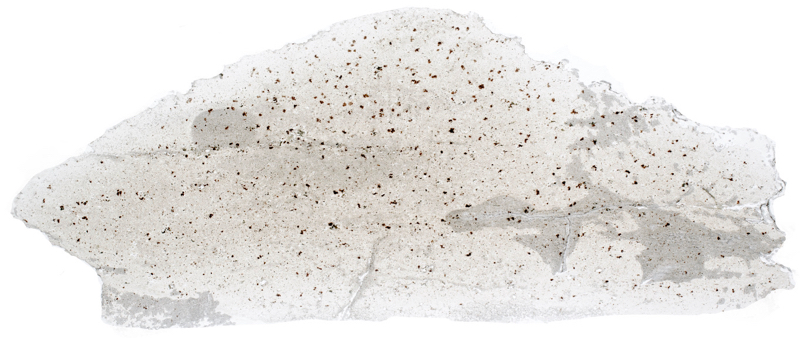
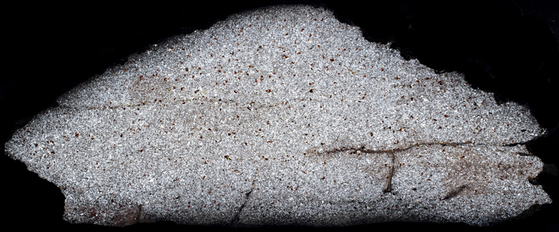
Location 5. Wester Hill south side rhyolite exposure at NT548315
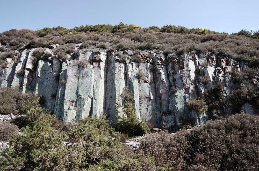
Guide books direct readers to a small disused quarry on the south-eastern side of the hill around the 300m contour where there is said to be ‘fine vertical jointing, of a columnar type’ in the felsite. A small quarry is marked on OS maps in this area but we find it to be completely full of rock fragments with no exposures visible. However, we do pass what could have been a small quarry on the south-western side of the hill where exposures display columnar jointing the rhyolite.
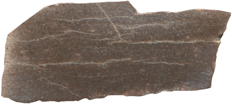
Prepared specimen viewed in plain reflected light (measures 45mm across)
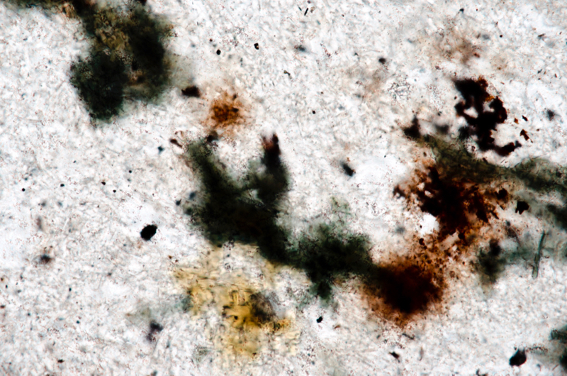
Section viewed in plane polarised light (FoV 0.5 x 0.3 mm)
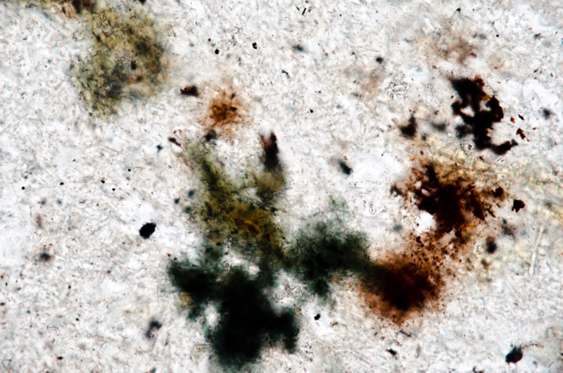
Section viewed in plane polarised light (FoV 0.5 x 0.3 mm)
Location 6. Mid Hill riebeckite-trachyte exposure at NT547323

We go down to the footpath at the base of Wester Hill and follow it around past Little Hill to the col between the Mid and Wester hills. We head up over the scree and along the side of Mid Hill to the outcrops visible from the col.
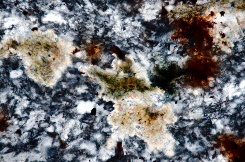
This rock and that at the summit is quite different from that on Wester Hill. It is of two types; riebeckite-trachyte and, at the summit, augite-olivine-trachyte. McRobert reports that specimens may be found that show the mixing of the two types.
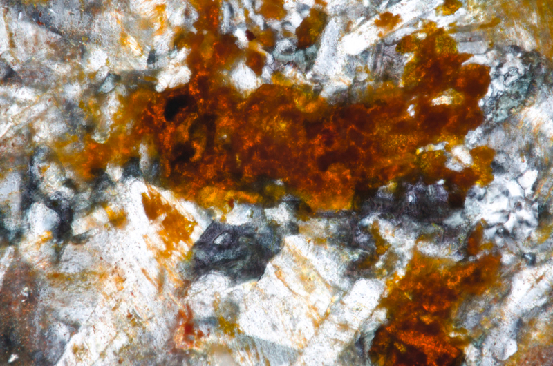
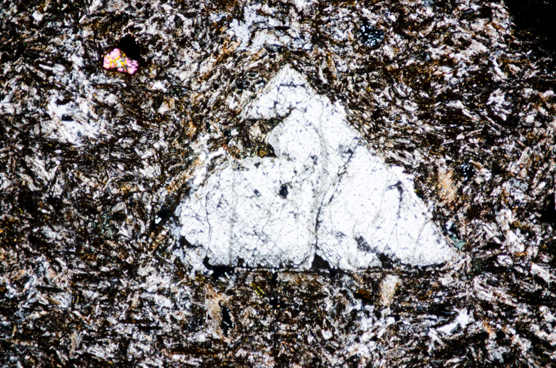
She describes the riebeckite-trachyte as being, ‘a very hard, compact, brown rock with a contorted fluxion-cleavage, recalling in appearance corrugated iron.’Under the microscope, ‘large and fairly abundant phenocrysts of anorthoclase lie in an orthophyric ground-mass of sanidine-prisms, embedded in deep-blue pleochroic riebeckite, and a small quantity of interstitial primary quartz.’ We are glad to see that these exposures seen from the col do contain riebeckite with its unmistakeable intense blue pleochroism.
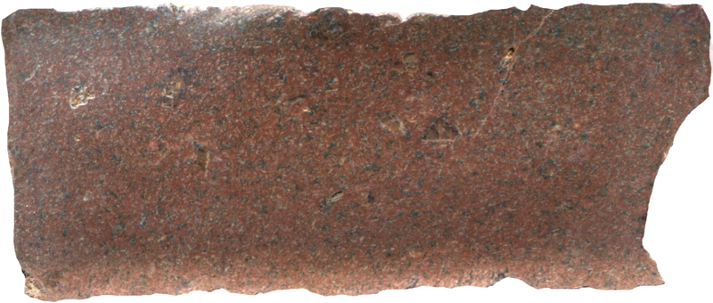
Prepared specimen viewed in plain reflected light (measures 42mm across)

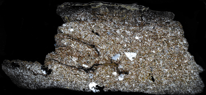
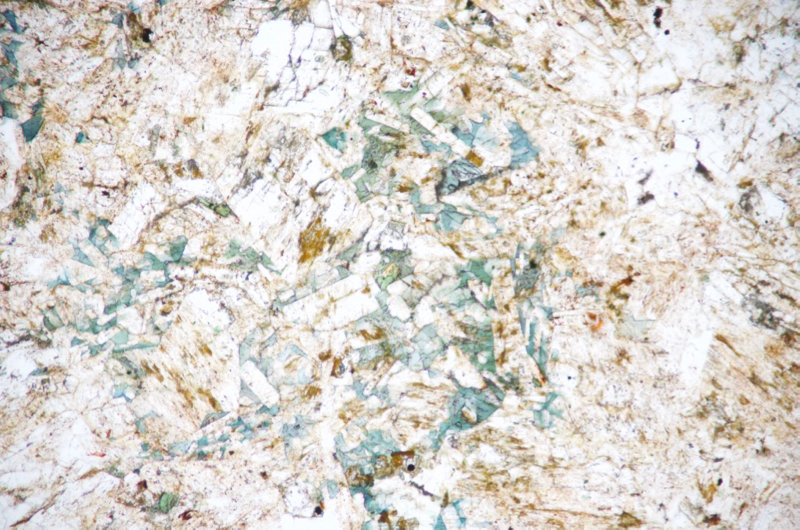
Section viewed in plane polarised light (FoV 1.2 x 0.8 mm)
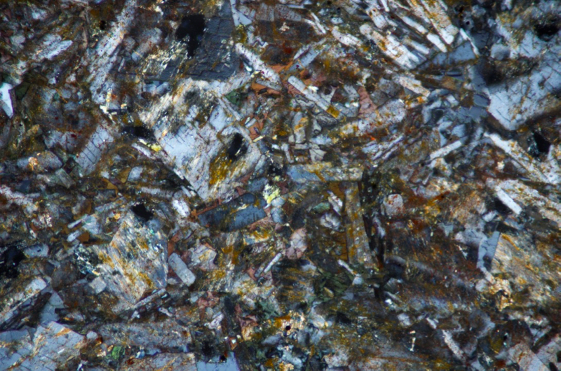
Section viewed with crossed polarising filters (FoV 1.2 x 0.8 mm)
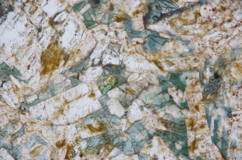
Section viewed in plane polarised light (FoV 0.5 x 0.3 mm)
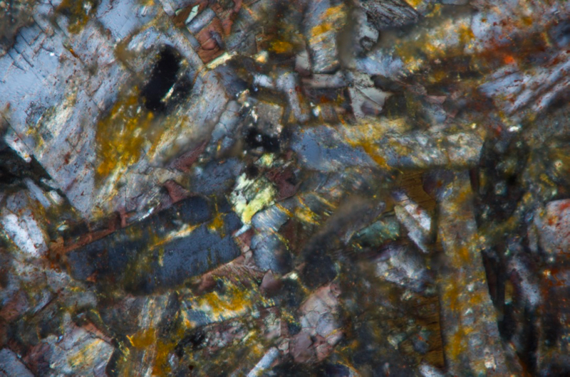
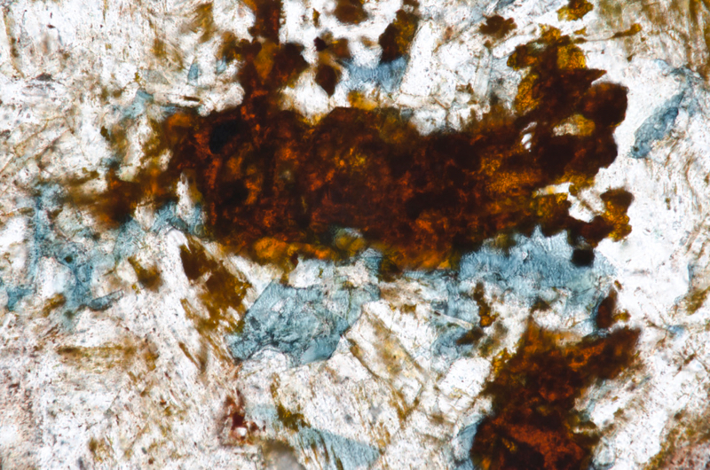
Section viewed in plane polarised light (FoV 0.5 x 0.3 mm)
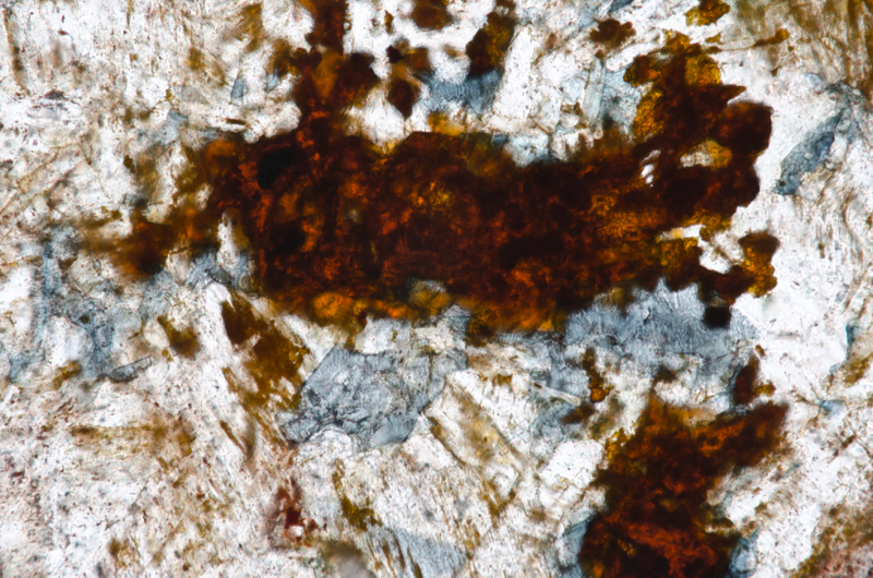
Section viewed in plane polarised light with polariser rotated 90deg.to show pleochroism (FoV 0.5 x 0.3 mm)
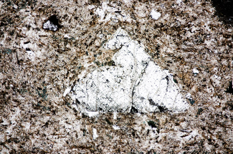
Section viewed in plane polarised light (FoV 4.6 x 3.0 mm)
Location 7. Mid Hill summit junction of rock types at NT547322
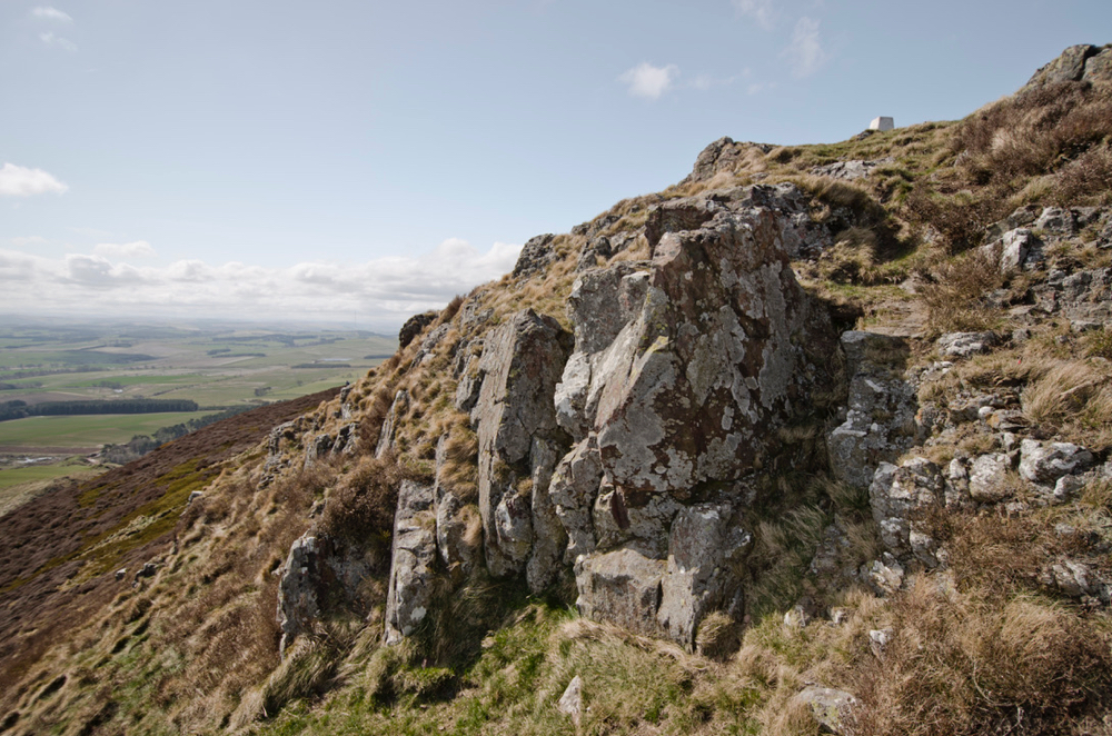
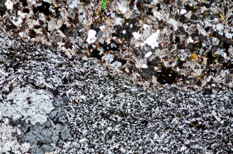
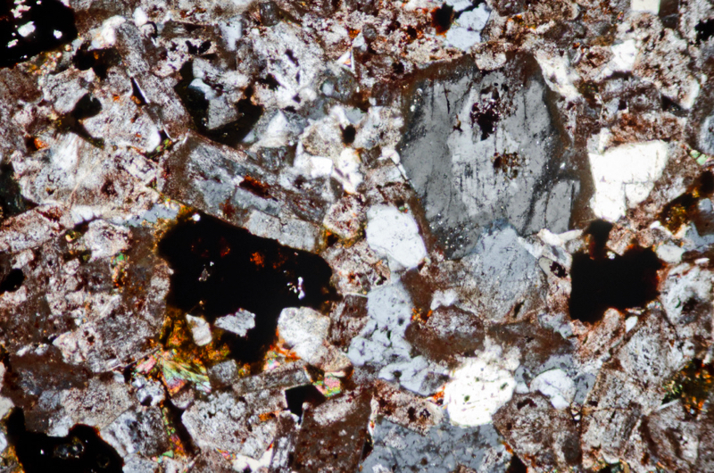
Just below the summit of Mid Hill, on the south side at NT547322, we find an interesting sample that is a mix of a dark-grey, finer-grained olivine-augite-trachyte and a red-brown, coarser-grained granitic-looking rock that may once have contained riebeckite (we see alteration products that may have originated in riebeckite) but looks more granitic than trachytic. We don’t see any olivine in the former, but we do see augite phenocrysts as well as albitised plagioclase phenocrysts. In the latter, along with alkali feldspar, ferromagnesian minerals and iron-titanium oxides, we see quite a lot of quartz.
Manson and Eckford’s 1927 paper, ‘A xenolith in riebeckite-trachyte, Roxburghshire’ described a xenolith found in the riebeckite-trachyte of Eildon Mid Hill that had a granitic character. They described the main constituents of the xenolith as orthoclase in euhedral stout prisms, quartz, aegerine, riebeckite, iron oxides with zircon as an accessory mineral and argued for the original presence of pyroxene on the grounds of suitably shaped pseudomorphs in quartz and iron-oxide.
They wrote, ‘An important feature of the xenolith is its drusy character’ – referring to the fluid-filled vesicles and fractures in the rock. They also noted the absence of chilling and interaction at the ‘quite sharp’ junction of the host and the xenolith.
We wonder if we have happened upon a similar mix of rock types.
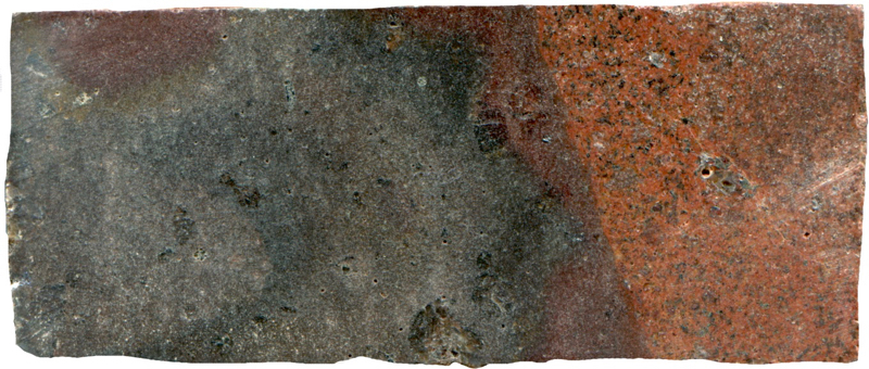
Prepared specimen viewed in plain reflected light (measures 42mm across)
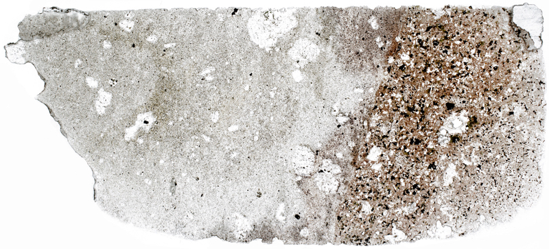
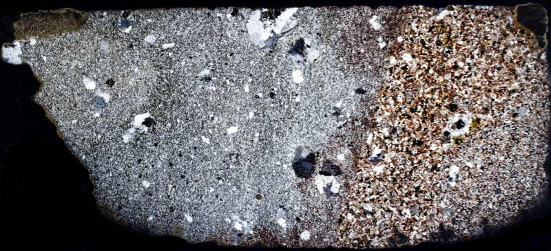

Section viewed in plane polarised light (FoV 4.6 x 3.0 mm)

Drawing by W. Manson, 1927

Section viewed in plane polarised light (FoV 1.2 x 0.8 mm)
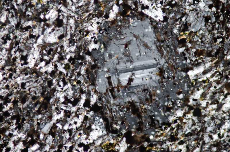
After alteration, only the central area of the plagioclase has retained its polysynthetic twinning (FoV 1.2 x 0.8 mm)
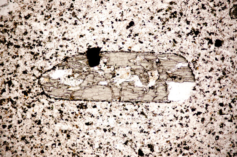
Portions of the crystal have disintegrated in the process of making the thin section. Section viewed in plane polarised light (FoV 1.2 x 0.8 mm)
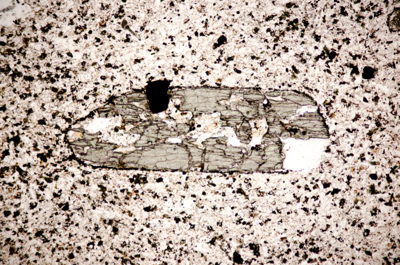
Section viewed in plane polarised light (FoV 1.2 x 0.8 mm)

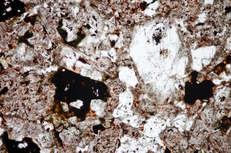
Section viewed in plane polarised light (FoV 4.6 x 3.0 mm)

Drawing by W. Manson, 1927.
Key:A. Late crystallised riebeckite (R) and iron-ore (black) moulded on felspar (F) faces.
B. Octagonal pseudomorph in iron-ore and quartz, probably after pyroxene enclosed in felspar.

This has the look of a ‘drusy’ portion of the rock – an area of alteration around a vesicle filled with crystallised hydrothermal fluid. Section viewed with crossed polarising filters (FoV 1.2 x 0.8 mm)
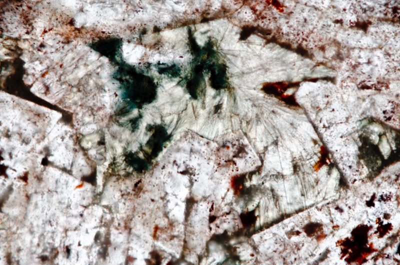
Section viewed in plane polarised light (FoV 1.2 x 0.8 mm)

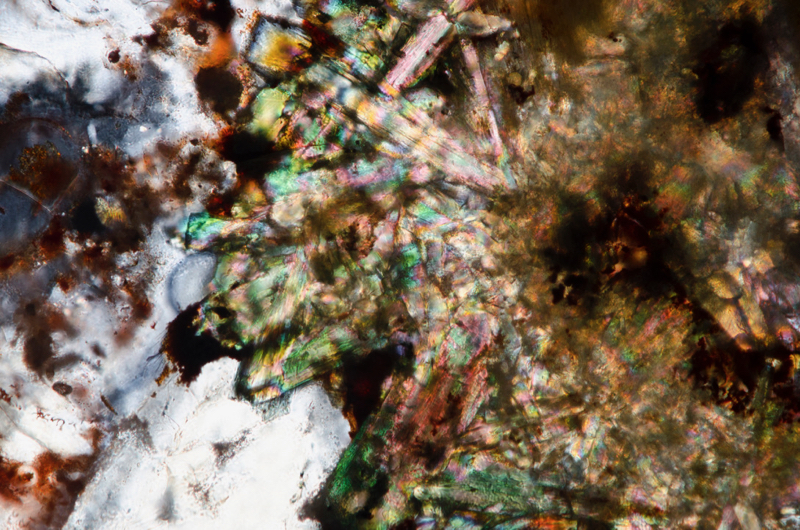
Section viewed in plane polarised light (FoV 0.5 x 0.3 mm)
Location 8. Mid Hill summit olivine-augite-trachyte at NT547322
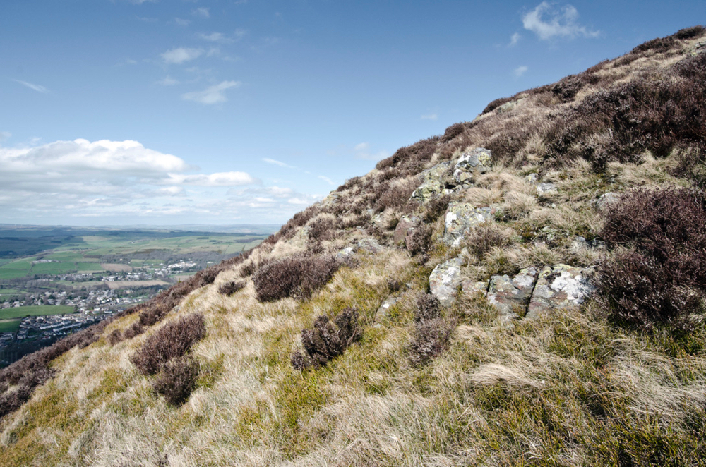
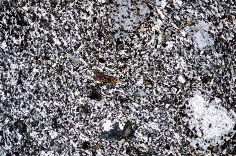
Around to the western side of the summit, we find small outcrops of augite-olivine-trachyte. Here, it is a dark, irregularly fractured rock with a greenish colour that weathers brown. Under the microscope we see orthoclase, tending to anorthoclase, and aegirine-augite phenocrysts in a trachytic sanidine and abundant, fresh, bright-green aegirine-augite groundmass. As in the sample we found earlier on the south side of the summit, the plagioclase phenocrysts have been albatised so they have lost all or most of their polysynthetic twinning.
What were occasional olivine crystals are now altered to characteristically shaped, yellow to red-brown pseudomorphs.
Lower down on this west side of Mid Hill, around the 210m contour, three tongues of the basal sanidine-trachyte cut across the Old Red Sandstone pedestal into the underlying Silurian rocks. These are understood to be feeder-dykes to the laccolith that would have been centrally situated beneath the structure; suggesting the original intrusion would have continued much further to the west to include the trachyte that is revealed at Bowdenmoor quarry.
First sample
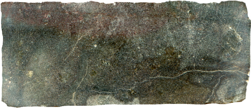
Prepared specimen viewed in plain reflected light (measures 42mm across)
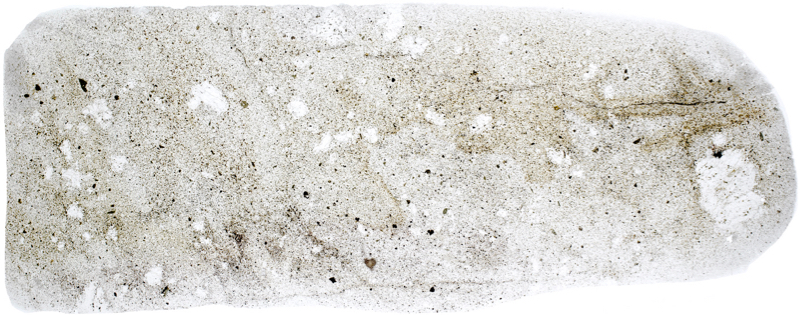
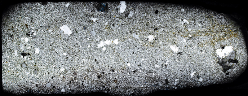
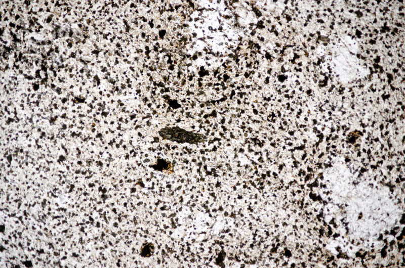
Section viewed in plane polarised light (FoV 4.6 x 3.0 mm)
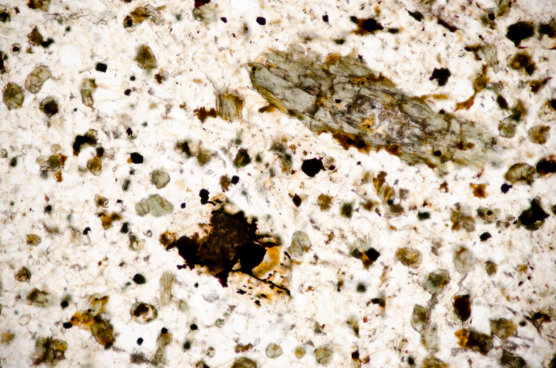
Section viewed in plane polarised light (FoV 1.2 x 0.8 mm)
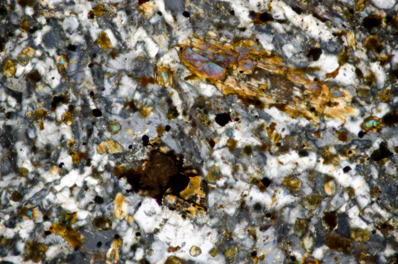
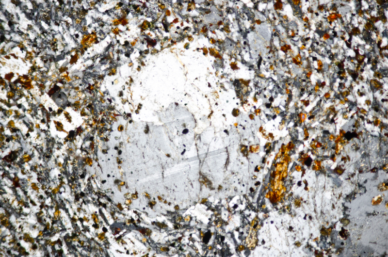
The remaining polysynthetic twinning is feint but visible in this phenocryst. Section viewed with crossed polarising filters (FoV 2.3 x 1.5 mm)
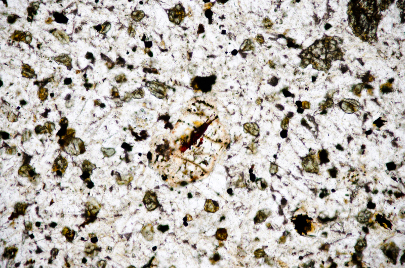
Section viewed in plane polarised light (FoV 1.2 x 0.8 mm)
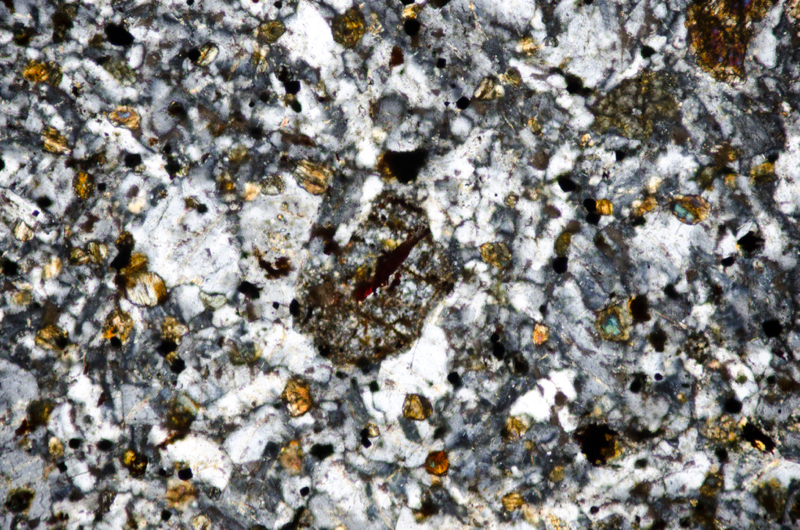
Second sample
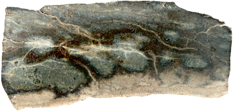
Prepared specimen viewed in plain reflected light (measures 42mm across)

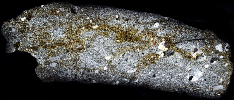
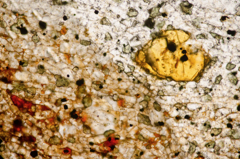
The alteration here has rendered the olivine a rich golden yellow. Section viewed in plane polarised light (FoV 1.2 x 0.8 mm)
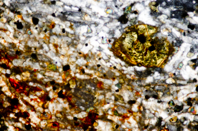
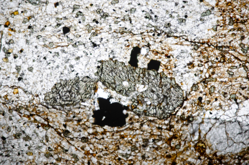
Section viewed in plane polarised light (FoV 2.3 x 1.5 mm)

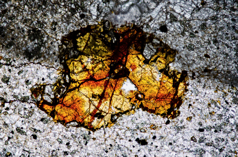
There are a few grey, non-yellow-orange, areas of the phenocryst that are relatively unaltered. Section viewed in plane polarised light (FoV 2.3 x 1.5 mm)
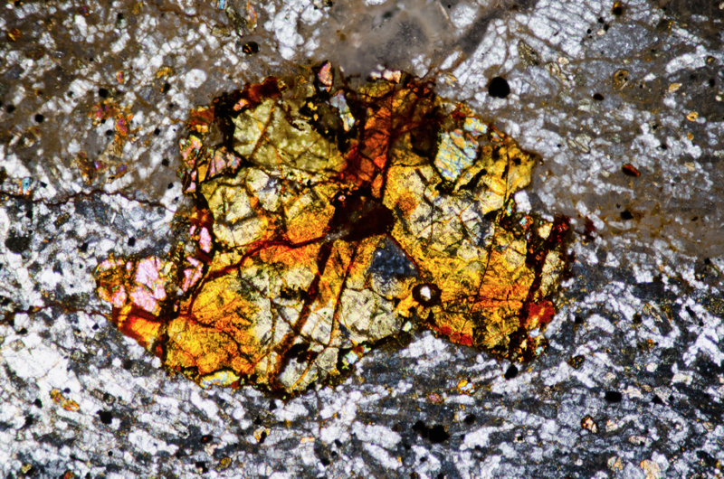
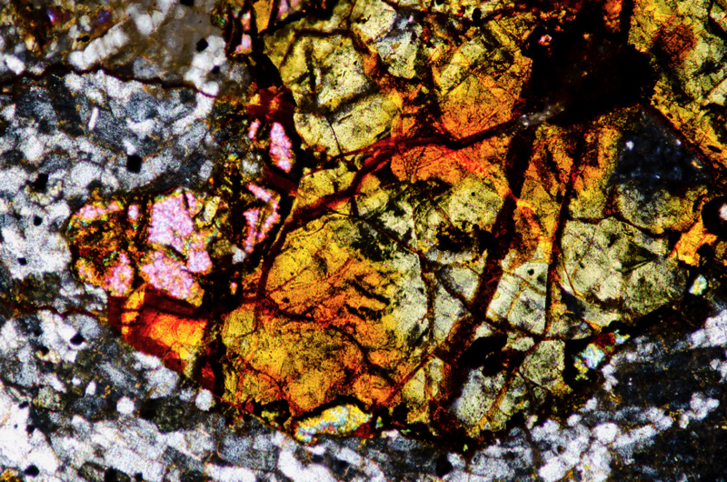
Section viewed with crossed polarising filters (FoV 1.2 x 0.8 mm)
Location 9. North Hill quarry sanidine-trachyte at NT550325
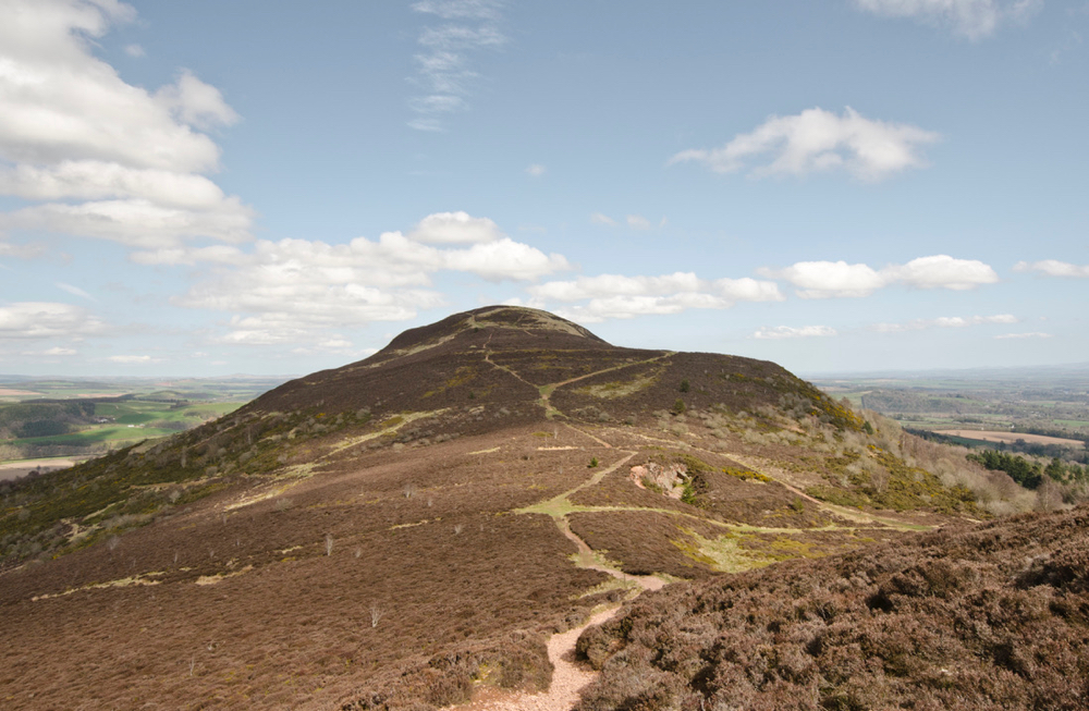
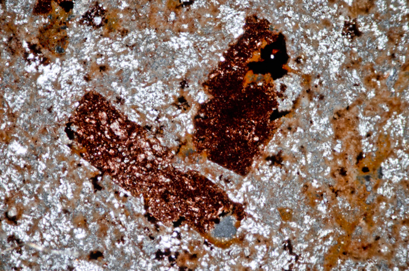
We follow the footpath north and down across to the disused old quarry at the bottom of North Hill. Here, between two sheets of basal sanidine-trachyte, there’s a thin bed of baked mudstone. Referring to the red, upper trachytic layer, McRobert noted, ‘the chilled edge against the mudstone is remarkable for its spherulitic structure.’
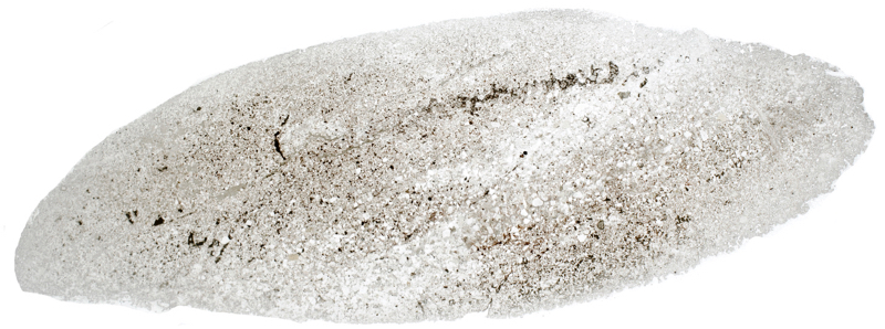
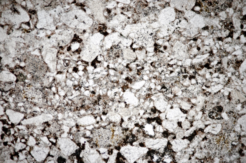
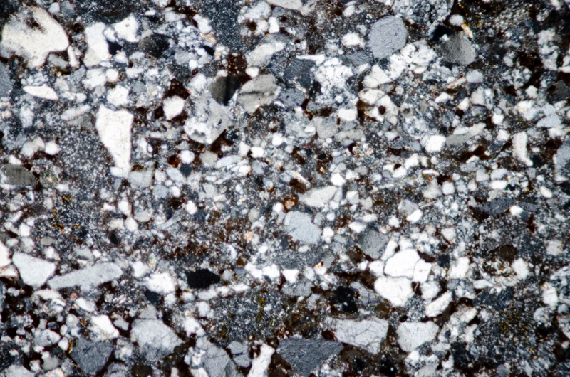
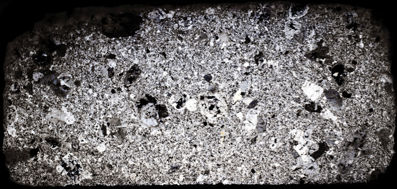
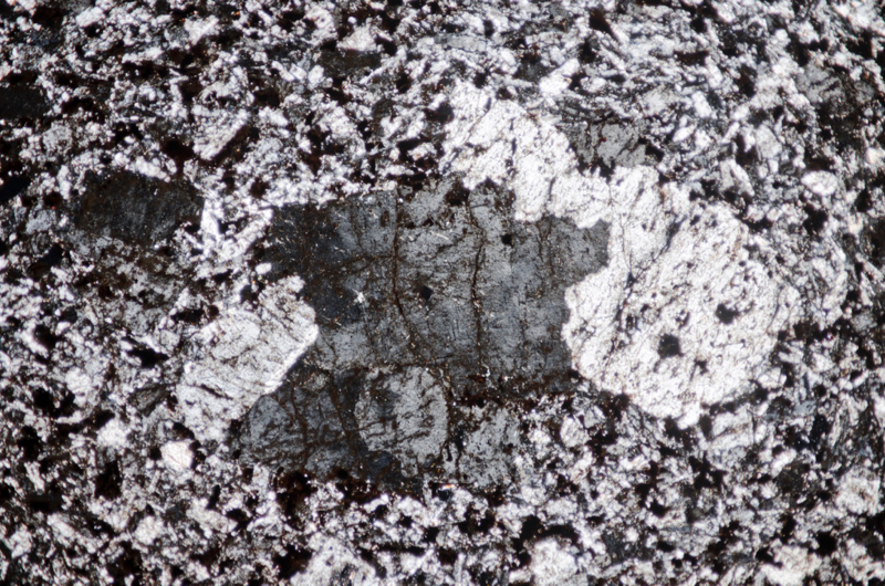
Our samples of sanidine-trachyte are very weak and their sanidine phenocrysts are cloudy or blackened by iron-titanium oxides. Our twin-tone sample of rock from the area where the mudstone meets the sanidine-trachyte turns out to be just mudstone, the change of colour being due to some areas containing more oxidised iron-titanium compounds than others.
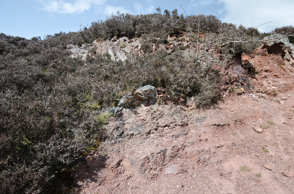
First specimen
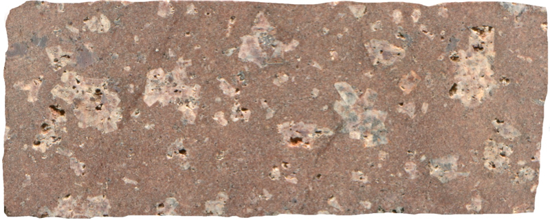
Prepared specimen viewed in plain reflected light (measures 42mm across)
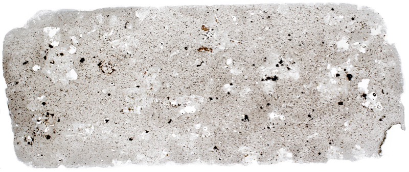

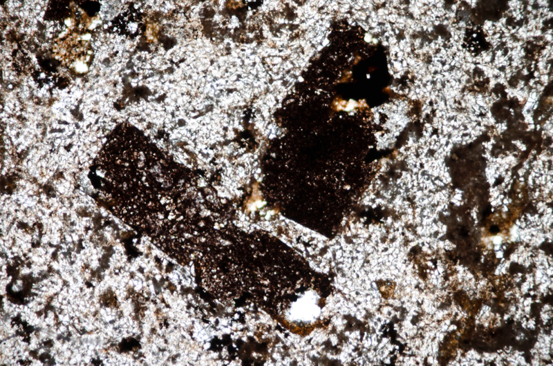
Section viewed in plane polarised light (FoV 2.3 x 1.5 mm)
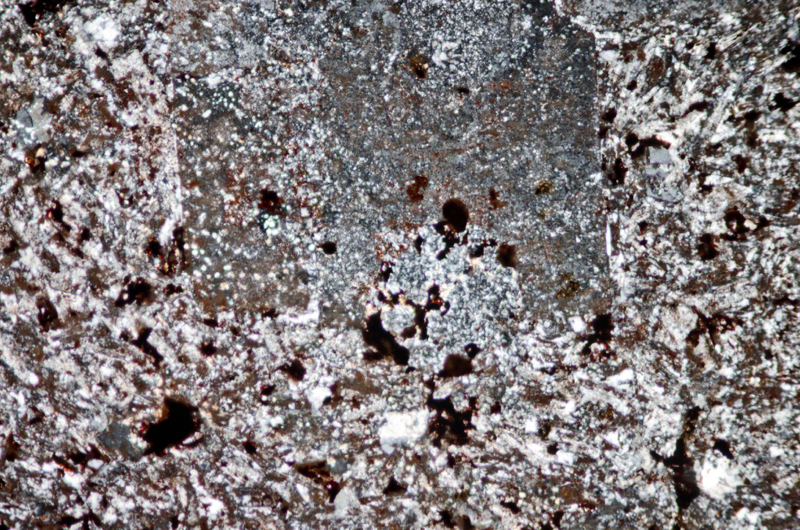
Section viewed with crossed polarising filters (FoV 2.3 x 1.5 mm)
Second sample

The portion of the rock richer in iron is more red as a result of its oxidation to haematite. Prepared specimen viewed in plain reflected light (measures 42mm across)
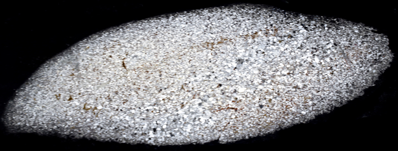
Section viewed in plane polarised light (FoV 2.3 x 1.5 mm)
Locations 10 & 11. North Hill summit sanidine-trachyte at NT554328
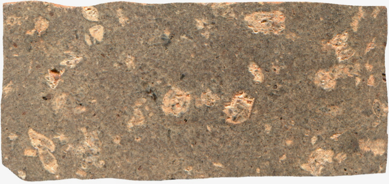
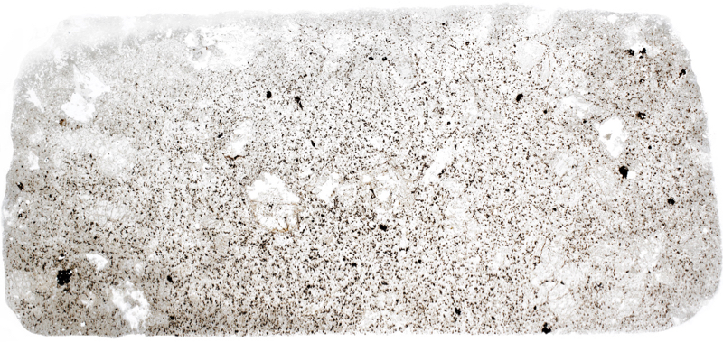
There are a couple of well-worn tracks up the grassy North Hill, we take the one at the eastern side of the quarry. Our walk to the summit is said to take us over three distinct layers of sanidine-trachyte. The lowest layer is the porphyritic type that we saw in the quarry lying on the Old Red Sandstone sedimentary rocks, a second, thicker layer, around location 10 on our map, that is reported as being quartz-rich but non-porphyritic, and then at the top of the hill, a highly porphyritic layer containing phenocrysts of fresh sanidine and euhedral quartz.
We see no definite outcropping rock on our way to the summit and so content ourselves, on this trip, with a sample from location 11, the summit. Here we appreciate the remaining ramparts of the Iron Age fort that later became the site of a Roman signal station. We also take in the views; south towards the volcanic hills at Minto, Peniel Heugh and Rubers Law, and north-east to White Hill and Black Hill with Great and Little Laws at Dirrington in the distance.
Prepared specimen viewed in plain reflected light (measures 42mm across)

Section viewed in plane polarised light (FoV 2.3 x 1.5 mm)
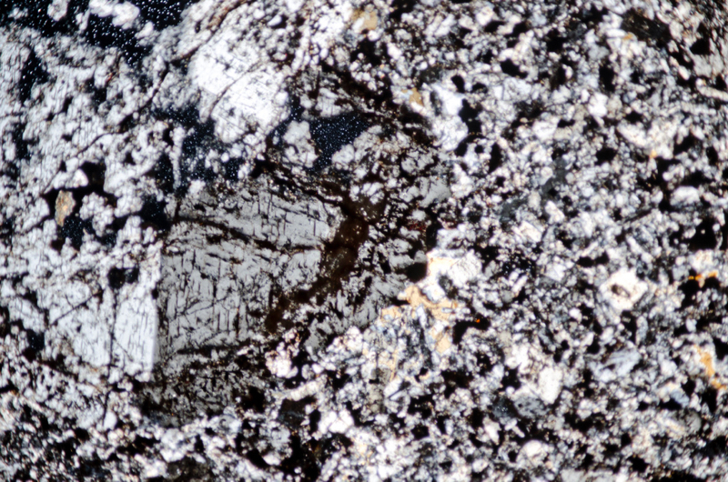
Section viewed with crossed polarising filters (FoV 2.3 x 1.5 mm)
References
R.W. McRobert, 1914, Acid and Intermediate Intrusions and Associated
Ash-Necks in the Neighbourhood of Melrose (Roxburghshire), Quart. Journ. Geol. Soc., Vol 70, pp.303-314.
W. Manson, R.J.A. Eckford, 1927, A xenolith in riebeckite-trachyte, Roxburghshire H.M. Geological Survey.
P. Stone et. al. 2012, British regional geology: South of Scotland.Fourth edition., Keyworth, Nottingham: British Geological Survey.
B.G.J. Upton et. al. 2003, Carboniferous and Permian magmatism in Scotland in Permo-carboniferous Magmatism & Rifting in Europe, M Wilsonet al. (editors), Geological Society.
N.H. Trewin, 2003, The Geology of Scotland, Geological Society; 4th revised edition.
A.D. MacAdam, E.N.K. Clarkson, P. Stone (editors) 1993, Scottish Borders geology: an excursion guide.Edinburgh, Scottish Academic Press.
BGS online Geology of Britain. http://mapapps.bgs.ac.uk/geologyofbritain/home.html